A picture is worth a thousand words. A picture is also not all that it seems. In chiropractic college, we
learn a lot about radiology, taking x-rays, reviewing said x-rays, as well as MRIs, CT scans, etc. We also
learn how to interpret and write reports on them. We are taught that 1 view is no view and you must
look at radiographs from at least 2 different angles. We are also taught not to rely solely on the findings
of the imaging because what’s on the image might not correlate all that well with a patient’s actual
symptoms.
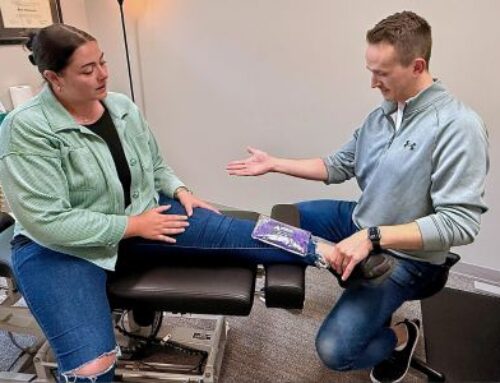
Let’s take a look at my recent MRI. As almost everyone knows, I suffered a severe, debilitating lumbar
spine injury, causing my intense radiating pain into my left hip and left lower leg. Upon getting the MRI
performed, I was fortunate enough to review the MRI in real time with one of my professors, who reads
advanced imaging, writes reports, and teaches others how to do it for a living. He took one initial glance
through my MRI and noticed something in the imaging that prompted him to say, “I’m assuming all your
symptoms are right sided”. Not at all.
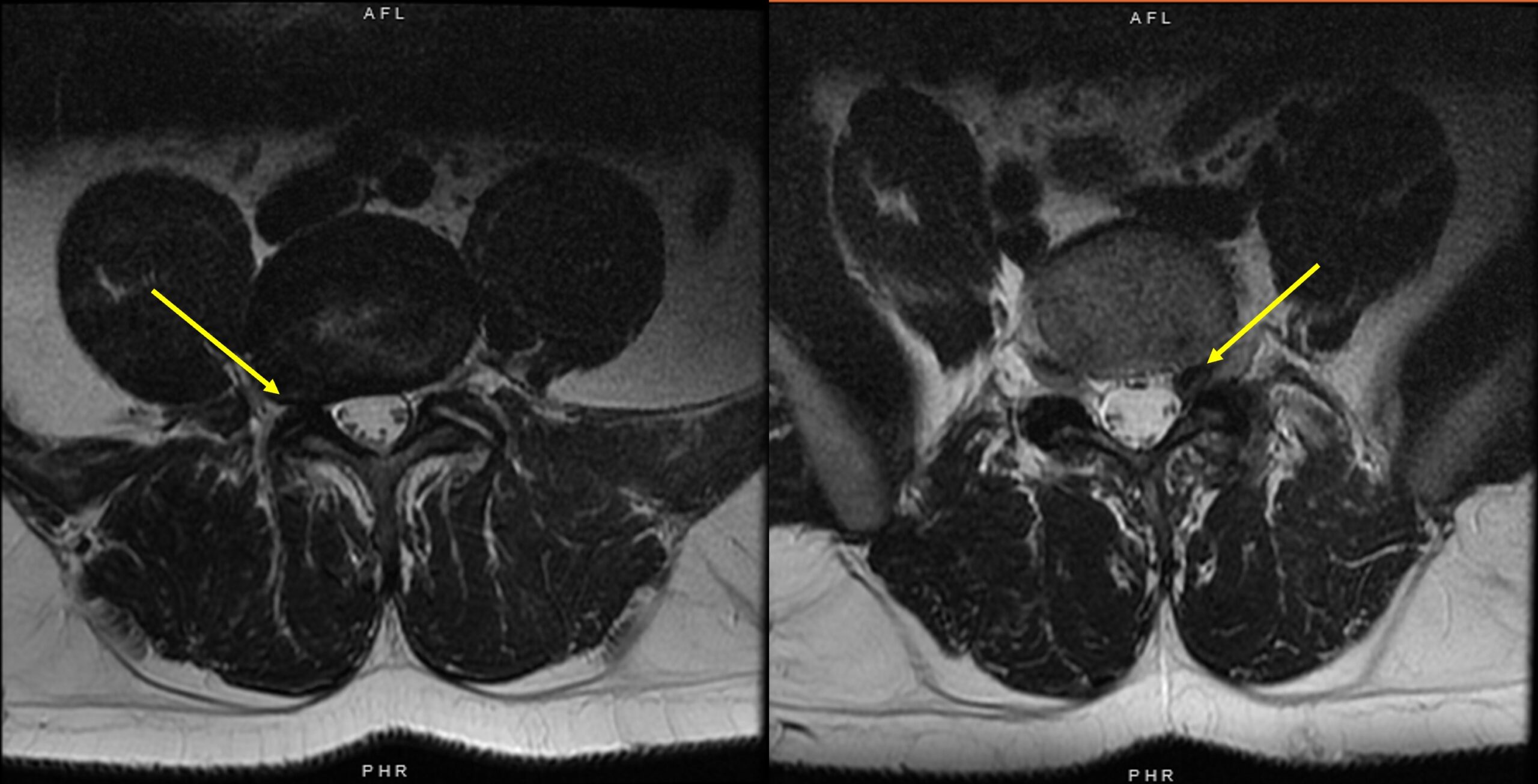
The reason he suspected that is because as your scroll through my MRI, from top to bottom, I have a
rather large and obvious disc protrusion on the right side 1 level above the problem area. And it’s large
enough to severely narrow the foramen, where the nerve exits the spinal canal. The image on the left is
the misleading image. This is the L4/L5 disc level (asymptomatic). The image on the right is the problem
area. This is the L5/S1 disc level, which you can only see on 2 MRI slices. It is not so obvious and a little
more inconspicuous.
I have never, in my life, had any right sided radicular pain, only left sided. This is true for when I suffered
a disc herniation in 2011, as well as what’s going on today. So, despite the fact that I have this massive
disc protrusion that severely narrows the foramen, it is completely asymptomatic at that location on
that side. This is why we take advanced imaging with a grain of salt. Sure, it provides a lot of good
information, but again, the findings on the image do not always correlate with the presentation of
symptoms.